
Retinal Detachment
What is Retinal Detachment?
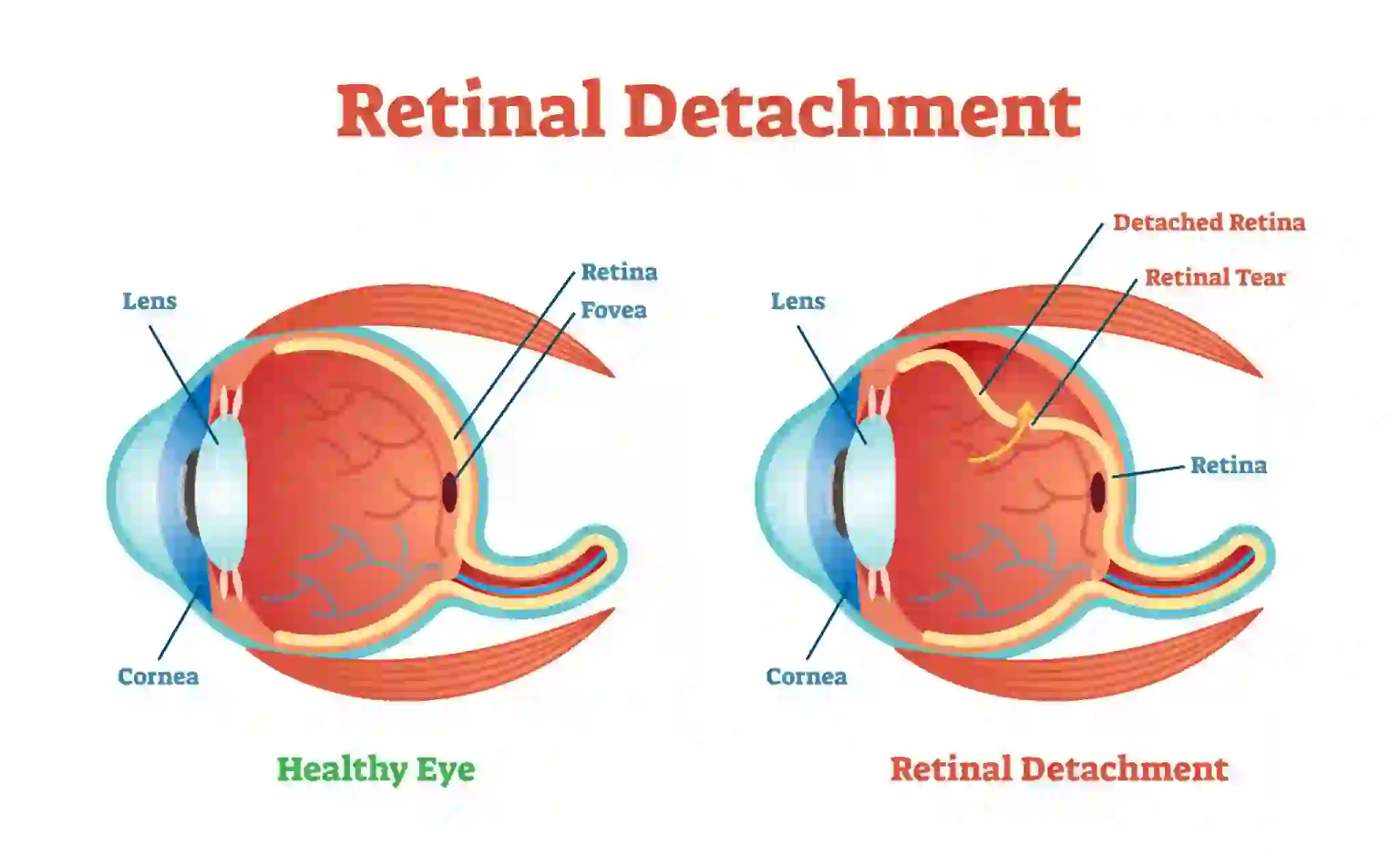
Retinal detachment is a serious eye condition in which the retina, the light-sensitive layer at the back of the eye, pulls away from its normal position. This separation prevents the retina from functioning properly, leading to vision impairment or blindness if left untreated. Common causes include aging, trauma, or underlying conditions like severe myopia. Early detection and prompt treatment are crucial to preventing permanent vision loss.
Retinal Detachment Symptoms
Recognizing the early warning signs of retinal detachment is crucial for timely medical intervention. Symptoms may develop suddenly or progressively, and they vary based on the extent and location of detachment. Below are some of the key indicators:
- Flashes of Light (Photopsia)
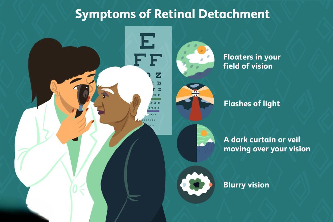
One of the most common symptoms of retinal detachment is experiencing brief flashes of light, known as photopsia. These flashes typically occur in the extreme peripheral vision and are unrelated to external light sources. They may appear as sudden, bright flickers, resembling lightning streaks. While occasional flashes can be harmless, persistent or increasing occurrences may signal a retinal tear or detachment.
- Sudden Increase in Floaters
Floaters are small, shadowy specks or thread-like shapes that move across your field of vision. While floaters are common with aging, a sudden and dramatic increase in their number could indicate retinal detachment. This happens when the vitreous gel inside the eye pulls away from the retina, sometimes leading to tears or breaks. Seeking immediate medical attention is recommended if new floaters appear alongside flashes of light.
- Ring of Floaters Near Central Vision
In some cases, people experience a concentration of floaters forming a ring-like pattern near the temporal side of their central vision. This symptom may be an early warning sign of a retinal tear before full detachment occurs. If left untreated, it can progress to severe vision impairment.
- Curtain or Shadow Over Vision
A significant symptom of retinal detachment is the appearance of a dark shadow or curtain descending over part of the visual field. This shadow may begin at the sides (peripheral vision) and gradually move toward the center, obstructing sight. It may also feel as though a veil is covering parts of the vision. This symptom usually indicates a progressing retinal detachment, requiring immediate medical intervention.
Appointment Making System
Our Eye care professionals will help you determine the most convenient time and schedule an appointment that suits your needs.
Book An Appointment- Veil-Like Effect in the Field of Vision
Another striking symptom is the impression of a veil or curtain being drawn over the field of vision. This may occur suddenly or progress gradually, worsening over time. The severity depends on how much of the retina is affected. If ignored, this can lead to irreversible blindness.
- Distorted Vision (Metamorphopsia)
Retinal detachment can cause straight lines to appear bent, wavy, or distorted. This distortion occurs due to the retinal layers shifting from their normal position, affecting how light is processed. People may struggle to read, recognize faces, or see fine details. If macular involvement occurs, distortion may be severe and permanent if left untreated.
- Loss of Central Vision
As retinal detachment progresses, central vision may become blurry or disappear entirely. This happens when detachment spreads toward the macula, the part of the retina responsible for sharp, detailed vision. The extent of vision loss depends on the severity and duration of detachment. If the macula becomes fully detached, surgery must be performed urgently to restore vision, though full recovery may not always be possible.
Causes of Retinal Detachment
Retinal detachment can occur due to various underlying conditions and risk factors. The most common retinal detachment causes include severe myopia (nearsightedness), ocular trauma, previous eye surgeries, and diabetic retinopathy. Identifying these risk factors early can help prevent retinal separation and protect long-term vision. Below are some of the major causes:
- Myopia (Nearsightedness) and Retinal Detachment
Severe myopia is a significant risk factor for retinal detachment. In individuals with high myopia, the eyeball is elongated, stretching the retina and making it thinner and more fragile. This increases the likelihood of retinal tears and lattice retinal degeneration, which can lead to retinal separation. Regular eye checkups are crucial for people with high myopia to monitor retinal health.
- Previous Cataract Surgery and Retinal Detachment
People who have undergone cataract surgery may have an increased risk of retinal detachment. During surgery, the natural lens is removed and replaced with an artificial intraocular lens (IOL). In some cases, this process can cause vitreous detachment, leading to retinal tears or exudative retinal detachment due to fluid accumulation. Patients who experience sudden retinal detachment eye flashes or floaters after cataract surgery should seek immediate medical attention.
- Ocular Trauma and Retinal Separation
Ocular trauma, including sports injuries, blunt force impacts, or accidents, can result in retinal detachment. A direct blow to the eye can cause the retina to tear or detach completely. Athletes and individuals in high-risk professions should use protective eyewear to minimize the chances of traumatic retinal detachment.
- Lattice Retinal Degeneration and Retinal Detachment
Lattice retinal degeneration is a condition where the peripheral retina becomes thinner and more vulnerable to tears. This degeneration is common in individuals with high myopia and can lead to spontaneous retinal detachment. Regular eye exams, including retinal detachment vision simulator tests, can help detect early signs of lattice degeneration and prevent serious complications.
- A Family History of Retinal Detachment
Genetics play a role in retinal detachment causes, as individuals with a family history of the condition are at higher risk. Certain inherited conditions, such as Stickler syndrome or Marfan syndrome, weaken retinal structures, increasing the chances of retinal separation. If there is a history of retinal detachment in the family, routine screenings are recommended to monitor retinal health.
- Diabetic Retinopathy and Retinal Detachment
Diabetes-related eye conditions, such as diabetic retinopathy, can lead to tractional retinal detachment. In advanced cases, abnormal blood vessels and scar tissue form on the retina, pulling it away from the back of the eye. This type of retinal detachment progresses gradually and may cause distorted vision, dark shadows, or central vision loss. Managing blood sugar levels and undergoing regular diabetic eye screenings can help prevent retinal separation.
Types of Retinal Detachment
Rhegmatogenous Retinal Detachment
What is Rhegmatogenous Retinal Detachment? This is the most common type of retinal detachment and…
What is Rhegmatogenous Retinal Detachment?
This is the most common type of retinal detachment and occurs due to a retinal tear or hole. The tear allows fluid to pass through and accumulate under the retina, causing it to detach. Risk factors include aging, high myopia, eye injuries, and previous eye surgeries. Flashes of light, floaters, and a dark curtain over vision are common symptoms.
Symptoms of Rhegmatogenous Retinal Detachment
- Very brief flashes of light (photopsia) in the extreme peripheral (outside of center) part of vision
- A sudden dramatic increase in the number of floaters
- A ring of floaters or hairs just to the temporal side of the central vision
- A dense shadow that starts in the peripheral vision and slowly progresses towards the central vision
- The impression that a veil or curtain was drawn over the field of vision
- Straight lines (scale, edge of the wall, road, etc.) that suddenly appear curved
- Central visual loss
Causes of Rhegmatogenous Retinal Detachment
Risk factors include the following:
- Myopia
- Previous cataract surgery
- Ocular trauma
- Lattice retinal degeneration
- A family history of retinal detachment
Rhegmatogenous Retinal Detachment Prevention
- Avoid direct and indirect injury to the eyes
- Regular eye check up
What is Rhegmatogenous Retinal Detachment?
This is the most common type of retinal detachment and occurs due to a retinal tear or hole. The tear allows fluid to pass through and accumulate under the retina, causing it to detach. Risk factors include aging, high myopia, eye injuries, and previous eye surgeries. Flashes of light, floaters, and a dark curtain over vision are common symptoms.
Symptoms of Rhegmatogenous Retinal Detachment
- Very brief flashes of light (photopsia) in the extreme peripheral (outside of center) part of vision
- A sudden dramatic increase in the number of floaters
- A ring of floaters or hairs just to the temporal side of the central vision
- A dense shadow that starts in the peripheral vision and slowly progresses towards the central vision
- The impression that a veil or curtain was drawn over the field of vision
- Straight lines (scale, edge of the wall, road, etc.) that suddenly appear curved
- Central visual loss
Causes of Rhegmatogenous Retinal Detachment
Risk factors include the following:
- Myopia
- Previous cataract surgery
- Ocular trauma
- Lattice retinal degeneration
- A family history of retinal detachment
Tractional Retinal Detachment
What is Tractional Retinal Detachment? This occurs when scar tissue on the retina pulls it…
What is Tractional Retinal Detachment?
This occurs when scar tissue on the retina pulls it away from the underlying layer. It is often linked to diabetic retinopathy, where abnormal blood vessels form and create tension on the retina. Over time, this traction leads to distorted vision and progressive vision loss.
Symptoms of Tractional Retinal Detachment
- Gradual diminution of vision
- Visual field defect which usually progresses slowly
- Straight lines (scale, edge of the wall, road, etc.) that suddenly appear curved
- Central visual loss if macula is detached
- Sudden drop in vision if associated with vitreous haemorrhage
Causes of Tractional Retinal Detachment
- Proliferative retinopathy due to diabetes
- Penetrating posterior segment trauma
- Vaso-occlusive lesions leading to fibrovascular proliferation
- Other causes such as retinopathy of prematurity, familial exudative vitreo retinopathy, idiopathic vasculitis
Prevention
- Controlling the systemic parameters such as blood glucose levels and blood pressure
- Regular eye check up
- Avoiding any trauma to the eyes
Types of Tractional Retinal Detachment
It can be classified based on type of vitreoretinal traction
- Tangential- caused by the contraction of epiretinal fibrovascular membranes
- Anteroposterior- due to the contraction of fibrovascular membranes extending from the posterior retina, usually in association with the major arcades, to the vitreous base anteriorly
- Bridging(trampoline)- due to contraction of fibrovascular membranes stretching from one part of retina to another or between vascular arcades
Diagnosis
- Ophthalmoscopy (direct and indirect ophthalmoscope)
- Fundus photography and Fundus Fluorescein Angiography
- Optical Coherence Tomography (OCT)
- Ultrasound B scan
Types of Rhegmatogenous Retinal Detachment
Long standing retinal detachment characterised by proliferative vitreo retinopathy changes
- Grade A- diffuse vitreous haze and tobacco dust
- Grade B-wrinkling of inner retinal surface & decreased mobility of vitreous gel
- Grade C- rigid full thickness retinal folds with heavy vitreous condensation and strands
- Ophthalmoscopy preferably with indirect ophthalmoscope
- Fundus photography
- Ultrasound B scan
- In the case of Rhegmatogenous Retinal Detachment Treatment, upon diagnosis, surgical intervention is the preferred choice for doctors in almost all instances.
- Retinal laser photocoagulation and cryopexy
- Scleral buckle surgery
- Pneumatic retinopexy
- Vitrectomy surgery
Tractional Retinal Detachment Treatment
- In the case of Tractional Retinal Detachment Treatment, upon diagnosis, surgical intervention is the preferred choice for doctors in almost all instances.
- Retinal laser photocoagulation
- Vitrectomy surgery
- Intravitreal anti vegf injections (bevacizumab, ranibizumab, aflibercept)
Sometimes a tractional retinal detachment can be stopped before it affects the central vision. A small area of retinal detachment far from the center of the vision can sometimes be watched if it stops growing due to retinal laser or anit vegf injection treatment and improvement in blood sugars control. Other times, a tractional retinal detachment affects the central vision significantly enough to require surgical repair. The surgery performed is called a vitrectomy, or removal of the jelly in the back of the eye that the abnormal vessels are growing into. Vitrectomy is also combined with careful microscopic dissection of the fibrous scars left by the abnormal blood vessels from the surface of the retina. Laser is often performed simultaneously to reduce the risk of the vessels recurring or to treat stretch holes in the retina. To help keep the retina reattached, the eye is sometimes filled with a synthetic gas or silicone oil at the end of the repair. Oftentimes, the decision to use one of those materials as a vitreous substitute is made during the surgery.
In conclusion, the of Tractional Retinal Detachment Treatment and other Eye Treatment is tailored to the individual’s needs and the severity of the condition. Early intervention, a comprehensive assessment, and collaboration between eye care professionals play a vital role in ensuring successful outcomes and improved visual function.
Retinal Detachment Prevention
Preventing retinal detachment is crucial, especially for individuals at higher risk due to factors like myopia, previous eye surgeries, or systemic conditions like diabetes. While not all cases of retinal separation can be avoided, the following preventive measures can help reduce the risk:
Avoid Direct and Indirect Eye Injuries
Eye trauma is a significant cause of retinal detachment, especially in people engaged in contact sports, hazardous jobs, or high-impact activities. To minimize the risk:
- Wear protective eyewear while playing sports like boxing, basketball, or racquetball.
- Use safety goggles when working in environments with flying debris or chemicals.
- Avoid rubbing the eyes aggressively, as excessive force can strain the retina.
- After an eye injury, seek immediate medical evaluation to rule out retinal tears or detachment.
Regular Eye Checkups
Routine eye examinations are vital for detecting early signs of retinal detachment, especially in individuals with high myopia, family history of retinal detachment, or pre-existing retinal conditions like lattice degeneration.
- People over 40 or those with risk factors should have an annual dilated eye exam.
- If experiencing retinal detachment eye flashes, sudden floaters, or vision distortions, consult an ophthalmologist immediately.
- Retinal detachment vision simulator tests can help patients understand potential vision changes and take preventive action.
Exudative Retinal Detachment
Unlike the other types, exudative retinal detachment is not caused by a tear but by fluid accumulation beneath the retina. Conditions like inflammatory eye diseases, tumors, or Coats’ disease can trigger this detachment. Symptoms may include blurry vision, visual distortions, and blind spots.
What are the Risk Factors of Retinal Detachment?
Here are some of the many risk factors of retinal detachment:
Several risk factors increase the likelihood of developing retinal detachment. While some people may have a genetic predisposition, others may develop it due to injuries or underlying conditions. Below are the key risk factors:
- History of Retinal Detachment in One Eye
People who have had retinal detachment in one eye are at a higher risk of developing it in the other eye. Regular monitoring and timely intervention can help prevent further complications.
- History of Eye Surgeries (e.g., Cataract Removal)
Individuals who have undergone cataract surgery or other intraocular procedures are more susceptible to retinal detachment. Surgical interventions can sometimes lead to vitreous detachment, increasing the chances of a retinal tear.
- Aging as a Risk Factor
Age-related changes in the vitreous gel inside the eye can contribute to retinal separation. As people age, the vitreous shrinks and may pull away from the retina, causing tears that lead to detachment. The risk is significantly higher after the age of 50.
- Severe Eye Injury and Retinal Detachment
Blunt trauma or penetrating injuries to the eye can cause retinal detachment by tearing the retina. Individuals involved in contact sports, high-impact activities, or accidents should take precautions to protect their eyes.
- Family History of Retinal Detachment
Genetics play a role in retinal detachment causes. If a close family member has experienced retinal detachment, the likelihood of developing the condition is higher. Routine eye checkups are essential for early detection.
- Myopia (Nearsightedness) and Retinal Detachment
People with high myopia (extreme nearsightedness) have elongated eyeballs, which stretch and thin the retina. This makes them more prone to lattice retinal degeneration and spontaneous retinal detachment.
- Underlying Eye Disorders and Diseases
Individuals with pre-existing eye conditions such as uveitis, lattice degeneration, retinoschisis, or Coats’ disease are at greater risk of retinal detachment. These diseases weaken retinal structures, making them more vulnerable to separation.
Controlling Systemic Risk Factors and Diseases (e.g., Diabetes)
Systemic conditions such as diabetes and high blood pressure increase the likelihood of tractional retinal detachment due to abnormal blood vessel growth. To minimize risk:
- Maintain optimal blood sugar levels to prevent diabetic retinopathy.
- Manage high blood pressure and cholesterol through lifestyle modifications.
- Follow a healthy diet rich in antioxidants and omega-3 fatty acids, which support retinal health.
When to See a Doctor
Knowing when to seek medical attention can prevent vision loss and ensure timely intervention. You should see an eye doctor immediately if you experience any of the following symptoms:
Emergency Signs of Retinal Detachment
- Sudden flashes of light (photopsia) in peripheral vision.
- A sudden increase in floaters, appearing like black spots or cobwebs in your vision.
- A dark shadow or curtain-like effect spreading across your vision.
- Distorted vision, where straight lines appear wavy or bent.
- Blurred or complete loss of central vision, especially if associated with eye conditions like exudative retinal detachment.
Routine Checkups Recommended If You Have
- High myopia (extreme nearsightedness), which increases the risk of retinal separation.
- A family history of retinal detachment, requiring preventive monitoring.
- Previous eye surgeries (e.g., cataract removal), which can lead to vitreous detachment.
- Diabetes or hypertension, increasing the risk of tractional retinal detachment.
- Ocular trauma, as even minor injuries can cause retinal damage over time.
While retinal detachment is a serious eye condition, early detection and preventive measures can help protect vision. Regular checkups, protective habits, and proper disease management are essential for those at risk. If you experience any warning signs like flashes, floaters, or vision distortion, consult an eye specialist immediately for evaluation and treatment.
Frequently Asked Questions (FAQs) about Retinal Detachment
Yes, eye flashes can be a warning sign of retinal detachment. They occur when the retina is being pulled or torn, leading to sudden, brief flashes of light in the peripheral vision. If accompanied by floaters or a shadow in vision, immediate medical attention is required.
With retinal detachment, vision may appear as if a dark curtain or shadow is covering part of the visual field. Other symptoms include sudden floaters, flashes of light, blurry vision, or a significant reduction in peripheral vision.
Early signs include sudden flashes of light, a sudden increase in floaters, blurry vision, or a dark shadow that spreads across the field of vision. If untreated, it can lead to permanent vision loss, so immediate medical attention is crucial.
Yes, a detached retina can occur again, especially if there are underlying risk factors such as high myopia, previous eye surgery, trauma, or weak retinal areas. Regular eye check-ups and following post-treatment care are essential to reduce the risk of recurrence.
- As mentioned above, there are several surgeries for retinal detachment like scleral buckle surgery, vitrectomy surgery, and pneumatic retinopexy. The last one is considered to be one of the simplest procedures to fix a retinal detachment. However, the only drawback is that it is not suitable in all cases.
- In the first step of this retinal detachment surgery, the surgeon carefully injects a gas bubble in the vitreous cavity of the eye to treat the damage or the tear with cryotherapy/freezing or laser. The injected gas bubble gently presses the retina of the eye against the wall of the patient’s eye, and the freezing or the laser slowly sticks the retina down, bringing it back to its original position. Lastly, once the retinal detachment surgery is over, it is crucial to take all the preventive measures to give some time for the injected gas to gradually disappear on its own.
The average cost for retinal detachment surgery in India is around Rs. 1,10,000. When it comes to health, it is always a good idea to invest in a good insurance plan that ensures that you do not have to go through a financial crunch in a time of need. On the other hand, there are several hospitals which will also allow you to pay the amount in installments according to your ease and convenience.
A scleral buckle is a type of retinal detachment surgery where the surgeon fixes a flexible, tiny band around the white area of the patient’s eye called the sclera. The role of this band is to gently push the sides of the eye while slowly moving it towards the retina to help the retina get reattached. Once this retinal detachment surgery is successful, the band will remain in the eye permanently to ensure there are no problems in the future.
After this retinal detachment surgery, most people are allowed to go home on the same day. However, they are asked to keep some pointers in mind like:
- Avoiding heavy exercises
- Wearing an eye patch for over a day.
- Having regular follow-ups with the doctor.
Also known as serous retinal detachment, exudative retinal detachment refers to a medical condition where fluid is collected behind the retina of the patient’s eye even though there are no tears or breaks in the retina.
- In this case, if a huge amount of fluid gets filled up, it can automatically push the retina away and cause detachment. Coats disease, trauma/injury to the eye, inflammation inside the eye, and age-related muscular degeneration (AMD) are some of the many causes of serous retinal detachment.
Usually, in most cases, retinal detachment surgery is required to treat this eye condition. It is imperative to understand that retinal detachment is an eye emergency that must be treated as soon as possible.
- The retina of the eye is similar to the film integrated into a camera. Therefore, in order to get a clear and accurate picture, it has to be smooth and healthy. In the surgery, the surgeon makes use of several medical tools and devices to ensure that the retina is fixed back in its place without causing any problems in the future.
Frequently Asked Questions (FAQs) about Rhegmatogenous Retinal Detachment
Yes, even a slight blockage of the vision caused by partial retinal detachment can result in blindness if not treated right away.
No. There is no medicine, eye drop, vitamin, herb, or diet that is beneficial to patients with retinal detachment.
Detachment is more likely to occur if the other eye has the condition (such as lattice degeneration) associated with retinal detachment in the first eye. If only one eye suffers a serious injury or requires eye surgery then, of course, the chance of detachment in the other eye is not increased by the event.
The outlook depends on the severity of the condition and how quickly you get expert medical care. Some people will recover completely, especially if the macula isn’t damaged. The macula is the part of the eye responsible for clear vision and is located near the center of the retina. However, some people may not regain full vision. This can occur if the macula is damaged and treatment isn’t sought quickly enough.
No. There is no medicine, eye drop, vitamin, herb, or diet that is beneficial to patients with retinal detachment.
Detachment is more likely to occur if the other eye has the condition (such as lattice degeneration) associated with retinal detachment in the first eye. If only one eye suffers a serious injury or requires eye surgery then, of course, the chance of detachment in the other eye is not increased by the event.
The outlook depends on the severity of the condition and how quickly you get expert medical care. Some people will recover completely, especially if the macula isn’t damaged. The macula is the part of the eye responsible for clear vision and is located near the center of the retina. However, some people may not regain full vision. This can occur if the macula is damaged and treatment isn’t sought quickly enough.
- In this case, if a huge amount of fluid gets filled up, it can automatically push the retina away and cause detachment. Coats disease, trauma/injury to the eye, inflammation inside the eye, and age-related muscular degeneration (AMD) are some of the many causes of serous retinal detachment.