Vascular endothelial Growth factor (VEGF) is a protein produced in the human body which is responsible for production of new vessels and maintaining them. Under abnormal conditions like diabetic retinopathy, blood vessels obstruction and age related macular degeneration it causes formation of abnormal vessels which bleed, leak and ultimately lead to scar formation and vision loss
What are anti VEGF Agents
Anti Vascular endothelial Growth factor (anti VEGF) agents a group of medications which block the activity of VEGF and thus mitigate the abnormal effects of VEGF
How are these anti VEGF agents different from one another
Bevacizumab
Ranibizumab
Aflibercept
Brolucizumab
Molecule
Monoclonal antibody
Antibody fragment
Fusion protein
Single chain antibody
Molecular weight
149 kDa
48kDa
97-115 kDa
26 kDa
Clinical dose
1.25 mg
0.5 mg
2 mg
6 mg
FDA approval
Not approved
Approved
Approved
Approved
Intravitreal anti VEGF activity
4 weeks
4 weeks
Upto 12 weeks
Upto 12 weeks
Appointment Making System
Our Eye care professionals will help you determine the most convenient time and schedule an appointment that suits your needs.
How have anti VEGF treatment influenced the management of various eye conditions
Anti VEGF agents when administered under appropriate conditions act at molecular level countering the action of VEGF and thereby reducing the morbidity.
Many diseases which were considered untreatable earlier like age related macular degeneration are rendered treatable, enabling patients maintain quality vision and subsequent improvement in quality of life
Ocular manifestation of systemic diseases with diabetes hypertension are also now treated with anti VEGF agents, with quality vision being restored and maintained.
What are the common conditions treated with anti VEGF agents and their benefits
Disease
Pathology
Pathology Benefits
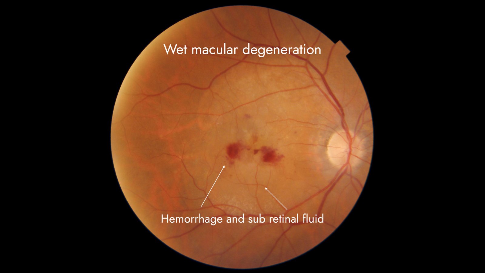
Wet age related macular degeneration
Abnormal vessels at the back of the eye leak fluid and blood, leads to drop in vision
Abnormal vessels regress with resorption of fluids with subsequent improvement of vision
Diabetic macular edema
Leakage of fluid due at the back of the eye leading to swelling and vision drop
Prevent leakage and reduce swelling
Proliferative diabetic retinopathy
Abnormal vessels on the retina which bleed
Regression of abnormal vessels
Retinal vein occlusion
Retinal vein occlusion Swelling of retina due to obstruction of retinal blood vessels
Resolution of swelling with improvement of vision
Disease
Pathology
Pathology Benefits
Wet age related macular degeneration
Abnormal vessels at the back of the eye leak fluid and blood, leads to drop in vision
Abnormal vessels regress with resorption of fluids with subsequent improvement of vision
Diabetic macular edema
Leakage of fluid due at the back of the eye leading to swelling and vision drop
Prevent leakage and reduce swelling
Proliferative diabetic retinopathy
Abnormal vessels on the retina which bleed
Regression of abnormal vessels
Retinal vein occlusion
Retinal vein occlusion Swelling of retina due to obstruction of retinal blood vessels
Resolution of swelling with improvement of vision
How do I choose the type of anti VEGF agent
The doctor examining you will prescribe the appropriate agents as per the disease process and systemic illness. Active bleeding or fluid leak at the back of the eye called macula warrants urgent treatment. The doctor will perform appropriate scans to confirm, quantify and monitor the progress of the disease. Vision is measured and is one of the yardsticks for monitoring response to treatment
What are the anti-VEGF agents available for treatment?
Bevacizumab
Ranibizumab
Aflibercept
Brolucizumab
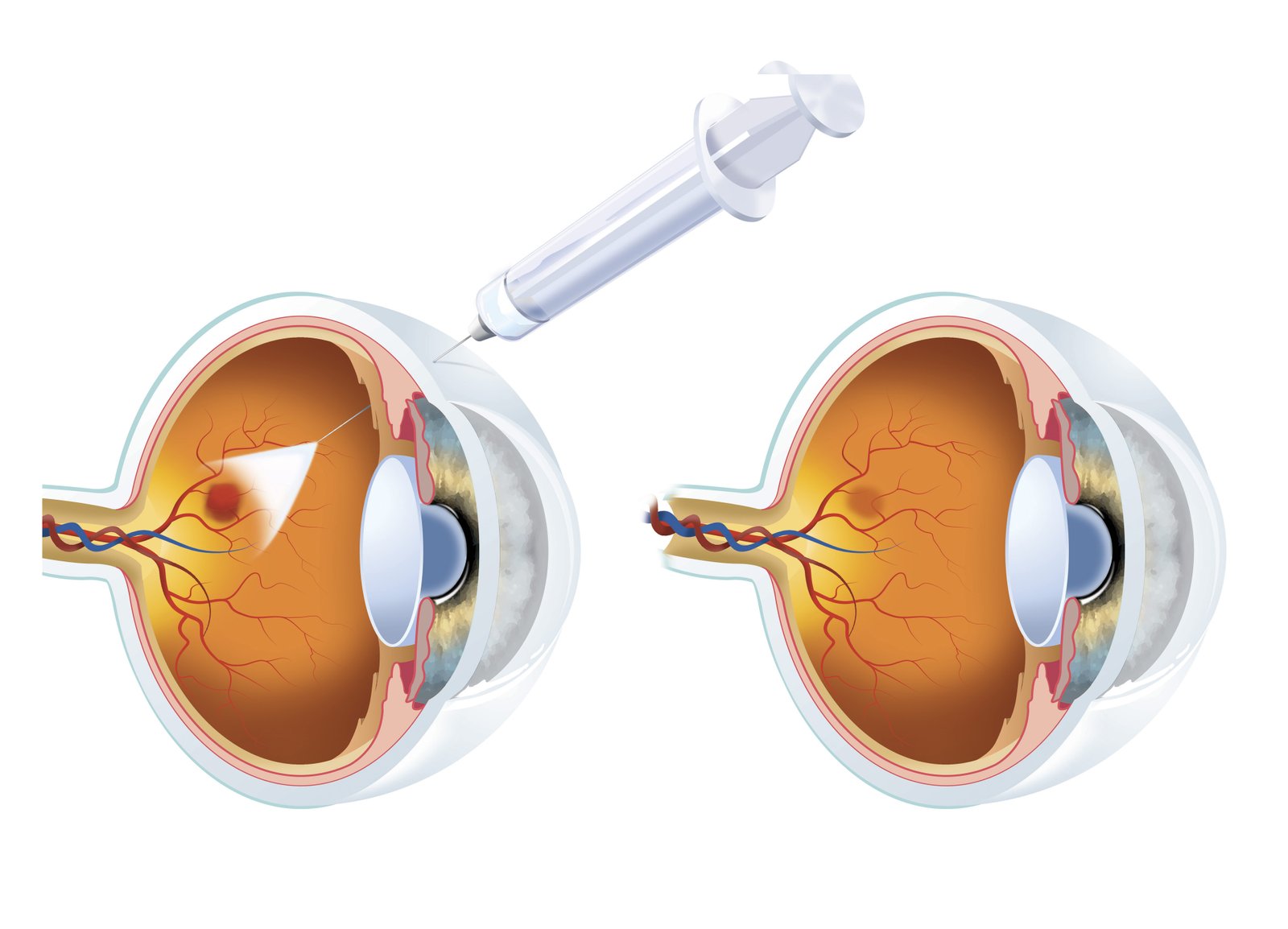
How is the Anti-VEGF agent administered
A macular hole is a small break or defect in the macula, the central part of the retina responsible for sharp, detailed vision. It can cause blurry or distorted central vision. Several factors can contribute to its development:
Primary Causes:
Aging (Vitreo-Macular Traction) – The most common cause. As we age, the vitreous gel inside the eye shrinks and pulls away from the retina. If it adheres to the macula too strongly, it can create a hole.
High Myopia (Severe Nearsightedness) – People with high myopia have thinner retinas, making them more susceptible to macular holes.
Trauma or Injury – Direct trauma to the eye, such as a blow or accident, can cause a macular hole.
Retinal Detachment or Epiretinal Membrane – Conditions that cause traction or pulling on the retina may lead to a macular hole.
Diabetic Eye Disease – Severe diabetic retinopathy can weaken the macula, increasing the risk of holes forming.
Macular Edema (Swelling of the Macula) – Fluid buildup can weaken the macular tissue, leading to hole formation.
Less Common Causes:
Inflammatory Diseases (such as uveitis)
Macular Telangiectasia (a rare retinal disorder)
Previous Eye Surgery (like cataract or retinal surgery, which can sometimes lead to macular holes)
Frequently Asked Questions (FAQs) about Anti VEGF agents
The chances of complications arising after the Anti-VEGF injections are extremely rare. Though most commonly, the problem arises from getting the injection in the eye, not the medicine. Some of the most common drawbacks are as follows-
Mild pain or ache in the eye can last two or three days
Floaters- will take a week minimum to get clear
Sclera may appear bloodshot or bruised
Eyes may feel rough, irritated, or puffy
These are common drawbacks of Anti-VEGF injections. However, if, in time, they don’t go away, then you should connect with your doctor and get a check-up done.
Bevacizumab injection is given to treat eye disease to block the abnormal growth of blood vessels in the back of the eye. The abnormal growth can block the vision and cause blood leaks in the eye resulting in vision loss.
The medicine takes approximately one month to show effect and improves vision. Though this depends upon your doctor and if they deem you fit for the eye injection. Patients with central retinal vein occlusion, myopic choroidal neovascularization, diabetic retinopathy, and other eye conditions are given bevacizumab injections.
The procedure is conducted inside the room and under the supervision of an experienced ophthalmologist. The surgeon might ask you to read a chart to check your vision. They will give eye drops to numb your eye, making the process painless.
Upon this, your eye will be cleaned with an ointment to prevent infection. Once done, the surgeon will place a tool to hold your eye open, or it will be hard to inject based on a human’s reflex mechanism.
Then the bevacizumab injection will be inserted in the sclera of your eye (the white part of the eye). The needle is extremely thin so as not to damage the eye or the vessels. The procedure will be painless, considering the numbing eye drops have been applied.
Once the procedure is over, the antiseptic and anaesthetics are washed from the eye and an eye patch is applied. Though an eye patch might not be mandatory, in a few cases, it is advised.
You are advised to consult with your ophthalmologist about what to do and what not to do. Please do not put any eye makeup on, refrain from straining your eye, and do not rub it unnecessarily, or the process might not occur because of eye irritation.
Though both are the most commonly used VEGF agents and have similar active molecule parts, bevacizumab and ranibizumab are different. The Avastin Bevacizumab is the anti-VEGF, whereas ranibizumab is an antibody fragment.
In the systemic circulation, the bevacizumab has an extended half-life compared to ranibizumab. But the latter one is said to have better retina penetration and higher affinity than the Avastin Bevacizumab.
Note that ranibizumab is a monoclonal antibody that slows the abnormal eye blood vessel growth and decreases the leak from these vessels. It falls in the category of vascular endothelial growth factor antibody. It stops vision loss and penetrates the retina to stop the growth.
The aflibercept injection helps in treating the age-related wet macular degeneration that causes the vision loss, or loss in seeing straight, causing discomfort in reading, driving, watching tv or other activities. The solution is injected into the eye sclera with a very thin needle. Once the proper dosage is injected, your eye will be cleaned. After the medicine takes effect, the vision loss will be restored, and you can read without discomfort.